THIS PAGE CONTAINS GRAPHIC IMAGES


Cosmetic Surgeries

This Shar-Pei’s condition is called entropion of the eye. The eyelashes were curled in causing abrasions on the cornea and a lot of pain. To correct this we took a very small trim of skin around the eye, and this pulled out the eyelashes from the eye.

Great results in the eye allowing the eyes to be normal and no longer have any treatments. The dog was no longer in pain as well!

Poor Pacino is a Miniature Pinscher. Glaucoma issues caused his eye to have painful ulcers and enucleation gave him full relief!


This 11 year old Shih Tzu named Bailey had a trauma injury to the head. The damage was so bad that the eye had to be removed.

Henry got into a fight through the fence and had his nose shredded. Reconstruction was done and healed well.

This bulldog had a condition known as screw tail. This is where the tail grows back into the body. It was removed giving relief from all the pressure and discomfort. Here we are postop.


Dental Issues

This is a German Shepherd who likes to chew rocks. He has worn his teeth down and major cavities have formed. This condition was very painful as all of his tooth roots were exposed and he had infection issues.

This is after many hours of doing root canals to all of his teeth.

Here is a mouth that has been neglected for several years resulting in severe tartar and gingivitis. They were cleaned and polished just like a human dentist would do.

This was the after picture of the dental cleaning and polishing.

Many of our patients are monkeys who become aggressive. It is not unusual for us to receive requests to do something about their canine teeth. As an alternative to pulling the teeth, which could result in fracturing the jaw or causing the mouth and teeth to move out of alignment, we do root canals. Root canals are performed on the canine teeth to reduce their size so that they are level with the rest of the teeth. This keeps the animal from causing as much damage with his bite, the mouth is preserved in a healthier state, and people are safer from injury.

We use the same materials and procedures that your dentist uses.

After the root canal has been drilled we use a dental acrylic; a UV light cures and hardens the material. The tooth is now safe and hard!

This is an x-ray of a Boston terrier who had an elongated soft palate. This caused swallowing and breathing problems.

These two pictures show the before and after of the soft palate reduction surgery.

In rodents and rabbits, the incisor teeth keep growing. Here are three different examples of overgrown or broken incisors. It is best to monitor the mouth and have teeth trimmed periodically.


Ear Crops

All of the long ears, which would be the Dobermans and Great Danes, have their ears wrapped to the top of their head to minimize bleeding. Then, they are put in their first rack the next day for five days. Everyone is sent home on an antibiotic and Rimadyl which is for pain and inflammation.

Dobermans and Great Danes

Dobermans

Great Danes

Boxers

Dobermans

Bully Pitbulls


Exotics

This is a Tamarin who was brought to us for a spay.

This Amazon tore the ligament in his leg. We reattached it and put him in a splint for four weeks. He had a full recovery.

This is a Tufted Marmoset. He tried to jump from his mother’s shoulder to the refrigerator. He did not make it, and ended up with a fractured tibia. We put a short IM pin in it to give support without being too heavy and overbearing since he weighed less than 3/4 of a pound. The splint was removed in six weeks and he was completely normal.

Sugar Gliders for neuters; this helps to tame them down.

Fox spays.

Examples of more neuters.

String can get caught around tails and tighten which then cuts off the circulation. Death and necrosis of the rest of the tail follows if the problem is not resolved. These show before and after the surgery.

Gastrointestinal issues in a Bobcat.

This is a tiger who ended up getting a stomach impaction.


Eye Surgeries

This was a severe corneal ulcer that has been stained to appreciate the depth and size of the ulcer. The surface of the eye has been scored to stimulate healing. Then the third eyelid is pulled across for support and acts like a sling while the eye is medicated. This often takes one or two weeks.

Ferrets can also develop severe eye infections and need to undergo a similar treatment.

Here’s a sugar glider with an eye infection.

This boxer mixed dog received this injury when he was hit by a car. His eye popped out of its socket. This condition is called Hyphema which means it is filled with blood. We were able to get it back into the eye socket and control the hemorrhage. He was able to see again with aggressive treatment.

G.I. Blockages and Bladder Stones

Here are some various urinary bladder stones that we find from time to time due to diet issues.

These are hair bands that were ingested and needed to be removed surgically.


Mass Removals

A very large fibrinous mass was removed from the foreleg of this dog, and a lot of skin was lost when the tumor was taken. I used a special technique and suture pattern to try and have enough skin to close the wound. There is a lot of concern when there is that much tension on the incision and skin causing a compromise in the blood supply for healing. The special technique allows a cushion and better circulation for healing. It obviously did very well as you can see in the last picture that was taken 3 ½ weeks post surgery.

Tumors on rats are very common! They make such cuddly pets we tend to see a bunch of these.

Tumors of the spleen are very concerning and hard to detect. If they get large enough to rupture, your dog will bleed out very very quickly. If your pet goes off appetite, becomes lethargic and especially is tender in the abdomen please call us ASAP.

This was a mast cell tumor on a young Doberman named Scarlett. All the margins were removed so that the mass will not reoccur in the nose.

A cancerous mass.


Orthopedic Surgeries

A 4 1/2 year-old cat with a humeral fracture. We repaired it using an intramedullary pin and cerclage wire. When the fracture is healed, the IM pin will be pulled, and the cat will be up and running.

This was a fracture of the right tibia. It is another example of an intramedullary pin being placed and closed prior to putting the leg in a splint. Healing went great and the dog was up and four-legged in six weeks.

This young owl had a wing injury and fractured her humerus. We were able to secure the fracture with a surgical stainless steel wire using a cerclage pattern, and she was fully healed in 6 weeks.

This young capuchin monkey had a metabolic bone disease issue. Metabolic bone disease is a concern with an endocrine issue or improper nutrition. It often leads to bone fractures because of the lack of calcium structure, or soft bones that grow improperly. Nutrition is extremely important in these cases.

This is a fracture of the left femur in a Pit Bull mix male dog. This radiograph was taken a little over a year after the surgery. The Intramedullary pin had been removed and the cerclage wire remains with a great healing at the fracture site.

This Pomeranian had a medial luxating patella. An issue where the knee and ligament do not line up straight so the knee cap tends to pop to the inside of the knee and out of the joint. Over time, the patella rubs in and out from the patella groove and that becomes a very shallow cradle. This is the groove that we have gone in and deepened and put back in the chondral bone to help deepen and stabilize the knee joint. You can see the straighter alignment after the surgery was done. Other stabilizers can be used if needed.

Here is a fox terrier who fractured his radius and ulna. An intramedullary pin was drilled into the radius and secured and then the proximal end of the radius was toggled over and secured to the IM pin. The tension of the muscles and support of a splint makes it perfect to heal with no secondary arthritic issues or other frustrations.

This is a humeral fracture from a very young boxer puppy. There was concern of putting an individual or a pin through a growth plate.

Using this technique, it stabilized the humeral fracture without causing a stunted growth of the humeral length or arthritis in the joints. The dog grew up to have a normal healthy life.

This is an 11-year-old German Shepherd with severe issues in his back - degenerative intervertebral disc disease. The inflammation has been going on so long that it is beginning to try to fuse together and stabilize itself. This causes compression and pain of the pinched IV disc pushing up into the spinal canal and pushing on the spinal cord. A lot of sharp pain in the back occurs and numbness in the rear leg area or back half of the dog.

Skyler is a 4 1/2 year-old cat whose left front leg was caught in the fan belt under the hood of a truck. It is common in the spring and winter to get up under the car engine for warmth and these injuries can be devastating. The fracture could have been repaired but there was a total loss of blood supply and nerve innervation.

One week post op up. He is happy and running around as if nothing had happened. Skyler has learned to compensate very well.

This is Falcor. He is a 12-year-old neutered, male basset mix. He had a cervical disc rupture, and this is soon after his surgery. Two examples of cervical disc recovery are included in the videos below.


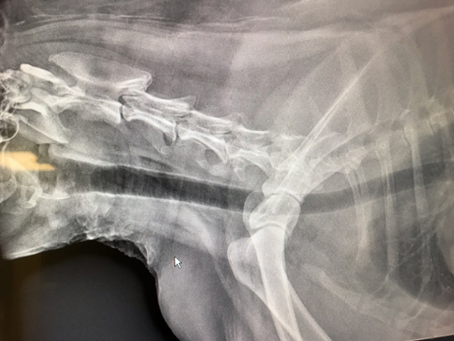
This next one is a seven year old malamute male that you saw in the second half of the video. He also had a cervical disc rupture. Looking at the x-ray you can see at cervical space 4/5 it has collapsed and calcified.

Here are 4 examples of dogs with Anterior Cruciate Ligament (ACL) injuries. These videos were shot within one to two weeks of post surgical repair.